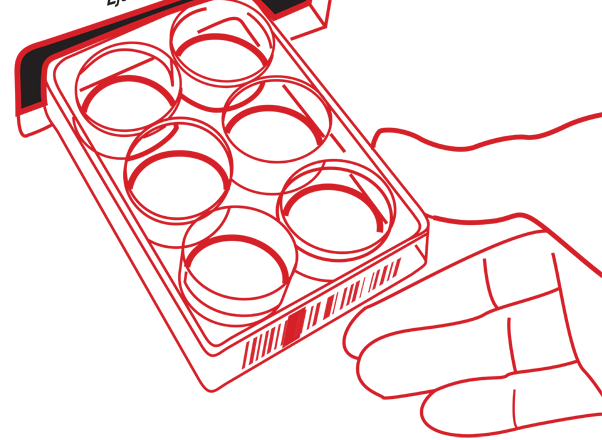
The CellAssist Software is at the heart of the CellAssist’s visualization and analysis capabilities. It can be placed in the laboratory or office to automatically download all of the images from the CellAssist instrument.
The CellAssist software analyzes the images, and presents a comprehensive and coherent view into all of the images and metrics from the entire history of all of the laboratory’s projects.
In addition, the entire plate setup information, normally entered into the CellAssist tablet, can also be entered into the software and automatically transmitted to the CellAssist instrument.
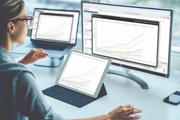
CellAssist Software improves quality and documentation of cells
Problem: Current tools do not collect images and data along with tools that enable users to visualize outliers. Outliers are important because they may indicate problems in quality that require investigation.
Objective: Document and analyze images and data on cells over time to improve quality and reproducibility of cell culture.
CellAssist Solution: CellAssist provides comparable data over time that allows for identification of outliers.
(In this example, upon further investigation, the customer found cases of mislabeling.)
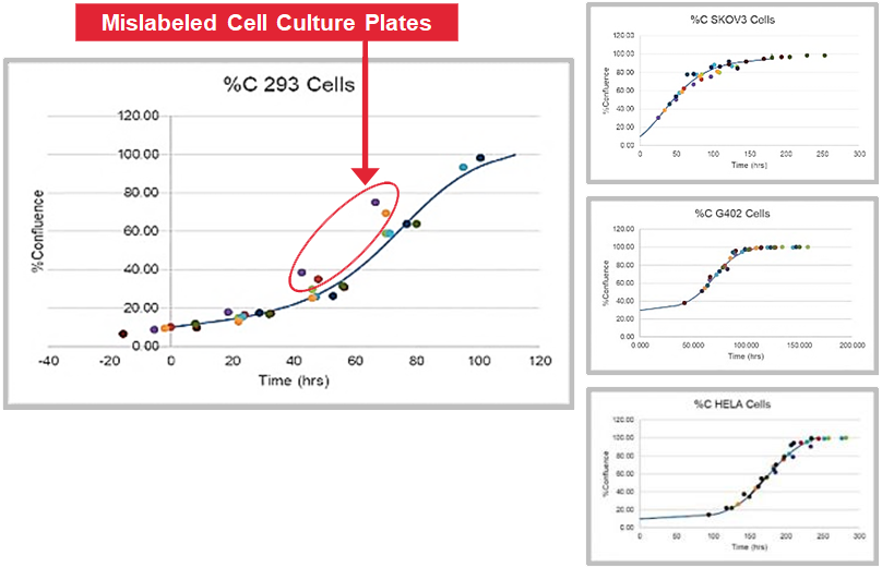
(Statistical anomaly highlighted)
CellAssist improves quality and increases yields by using data to grade and pick colonies
Problem: Gene editing transfection rates can be poor, so it is important that monoclonal colonies be selected for passaging.
Objective: Accurately determine whether a colony is monoclonal by capturing series of time lapse images with excellent registration across scans.
CellAssist Solution: The CellAssist time-lapse imaging enables the lineage of candidate colonies to be tracked to a single starting cell.
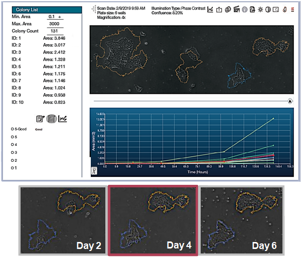
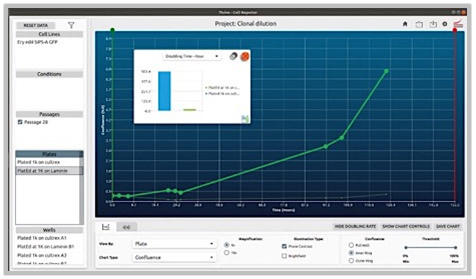
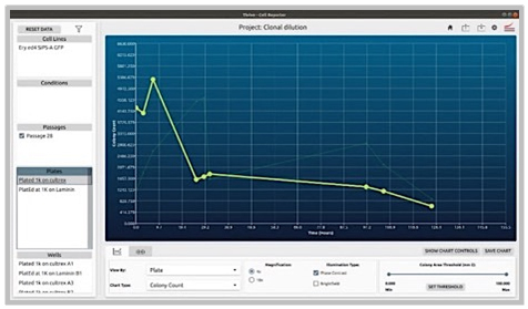
Problem: (a) The coating of plates can profoundly affect the survivability and growth of colonies (in this case gene edited ones starting with very small quantities). (b) Some coatings are also significantly more expensive.
Objectives: Compare two coatings to choose the optimal one by characterizing and comparing the cells under both conditions over time. (b) Tailor, test and directly compare the impacts of new protocols to select the best coating AND protocol.
CellAssist Solution: The CellAssist’s documentation, colony counting curves and examination of images are used to characterize the number of colonies for each condition, and the results are collated and easily compared across conditions.
Centralized database provides consistent and shareable images
to improve your cell, tissue and suspension culture
experiments and results
Objective metrics and quantitative reports
enhance cell, tissue and suspension culture
decision making and productivity
Key workflow details and status of cells are captured and archived
to provide confidence that data is not lost or compromised